Table of Contents
- 1What Is the Anatomy of a Large Bowel?
- 2What Is the Function of Colon?
- 3What Are the Types of Polyps?
- 4What Are the Benefits of Screening?
- 5What Are the Diagnostic Options For Colon Abnormalities?
- 6What Is the Procedure For MRI Colonoscopy?
- 7What Are the Types of MRI Colonoscopy?
- 8What Is Conventional Colonography?
- 9Which Is Better Among, MRI and Conventional Colonoscopy?
Introduction:
Malignant tumors in the large bowel are the second most common type of cancer. The tumors develop from long-standing adenomatous polyps. The cancer is diagnosed late due to challenges in screening tests because of their low sensitivity, poor detection of polyps, or high cost. The present-day screening includes MRI (magnetic resonance imaging) colonoscopy along with conventional colonoscopy to detect polyps.
What Is the Anatomy of a Large Bowel?
It comprises the colon, rectum, and anus. The entry point of the colon is called the caecum. The colon is divided into parts: the ascending colon (travels up), transverse colon (travels towards left), and descending colon (travels downward) and is followed by the sigmoid colon (travels back to the right side). The rectum is a fixed terminal part of the large intestine that is in continuation from the sigmoid colon. The inferior part of the rectum continues to the anal canal. The movements of the colon are segmentation contraction and peristaltic waves.
What Is the Function of Colon?
-
Enables the absorption of water, sodium, and other minerals from the chyme.
-
Aids in the conversion of isotonic chyme to semi-solid feces.
-
Contractions mix the content entering the colon and expose it to mucosa for absorption.
-
The peristaltic movement pushes the feces toward the rectum.
What Are the Types of Polyps?
The polyps appear sporadically in the colon and show an increased frequency with age.
Polyps can be of two types:
1. Non-neoplastic Polyps- It shows minimal cancer potential.
2. Adenomatous- A significant number of colon and rectal cancers originate from adenomas.
The non-neoplastic polyps include:
1. Hyperplastic Polyps- It is a common type of polyp caused by reduced epithelial cell turnover and accumulation of mature cells on the surface.
2. Hamartomatous Polyp- It is caused by a malformation in the stroma and appears sporadically or in association with genetic defects.
3. Inflammatory Polyp- Appears as inflamed regenerative mucosa surrounded by ulcers. It is seen in inflammatory bowel disease.
4. Lymphoid Polyp- It contains intramucosal lymphoid tissue and is a normal variant of the mucosa.
What Are the Benefits of Screening?
-
Early screening of pre-cancerous polyps can prevent their advancement to cancer.
-
Early detection of cancerous lesions can improve the long-term survivability of patients.
What Are the Diagnostic Options For Colon Abnormalities?
-
Double-contrast barium enema.
-
Conventional colonoscopy.
-
2-dimensional computed tomographic colonography (2D-CTC).
-
3- dimensional colonoscopy (3D-CTC).
-
3D Magnetic resonance colonography (MRC).
High radiation exposure is a limitation of using CTC.
What Is the Procedure For MRI Colonoscopy?
It utilizes fast sequence imaging and surface coil. Water or saline can be used as a positive contrast to distend the colonic lumen.
-
The patient is asked to hyperventilate.
-
In patients unable to hold respiration for 30 seconds, the turbo-spin echo technique is used to shorten the apnea duration.
-
Bowel preparation is done with macrogol 3350.
-
Intravenous administration of 5 mg (microgram) of scopolamine is made before the examination. It optimizes colon distention, minimizes peristaltic defects, and alleviates spasms.
-
The patients are made to lie in a prone position on the MRI table. The patient's colon is filled with 1,500 to 2,000 ml (milliliter) of water and 15-20 ml of 0.5mmol/L (millimoles per liter) gadopentetate dimeglumine solution.
-
The colon is monitored while filling with echo imaging.
-
When the contrast dye reaches the cecum, the MRI sequencing images are taken.
-
The imaging is done for supine and prone positions.
Advantages
-
Evaluation of small bowel by MRI in patients with inflammatory bowel disease is a new standard.
-
The scan demonstrates high sensitivity and specificity in detecting extraluminal lesions like abscesses and fistulas.
Limitations.
- Obvious colorectal lesions can be identified, but the images can be unsatisfactory despite the bowel preparation and distention status.
What Are the Types of MRI Colonoscopy?
Based on the signal in the lumen there are two types of MRI colonoscopy.
Bright lumen MRI colonoscopy
-
The colorectal lesions appear as dark filling defects with a bright lumen.
-
The effect is achieved with rectal enema containing paramagnetic contrast.
-
The contrast containing the colon lumen appears bright but the colon wall and polyp have low signal intensity.
-
The technique is less sensitive in differentiating between polyps, air, and residual stool.
Dark lumen MRI colonoscopy
-
It focuses mainly on the colonic wall.
-
The colonic wall is brightly enhanced, but the colonic lumen is homogeneously dark.
-
Only water is used as a medium rectally.
-
The colon filling process is monitored with fluoroscopy.
-
The paramagnetic contrast medium is administered intravenously.
-
Residual air does not produce a signal in the lumen. Therefore, the examination is carried out in a prone position only.
-
The polyps appear enhanced and, therefore, they are differentiated from the residual stool.
-
Lesions below 5 mm are difficult to identify in this technique.
-
Lesions above 8 mm are easily identified.
-
Additional polyps in the region that are difficult to identify through colonoscopy are diagnosed.
What Is Conventional Colonography?
-
The current gold standard for detecting colorectal polyps and inflammatory bowel disease.
-
It is a reliable technique for identifying colorectal cancer and differentiating between colorectal lesions.
-
There is an option for obtaining a biopsy, polypectomy, or treatment during the examination.
-
A flexible tube is inserted for the entry of a tiny camera to visualize the colon.
Limitations
There is a risk of observer error and the regions of the colon not screened with colonography prevent identifying polyps that are less than 5 mm in diameter.
It is not suitable for identifying obstructing colonic lesions.
Which Is Better Among, MRI and Conventional Colonoscopy?
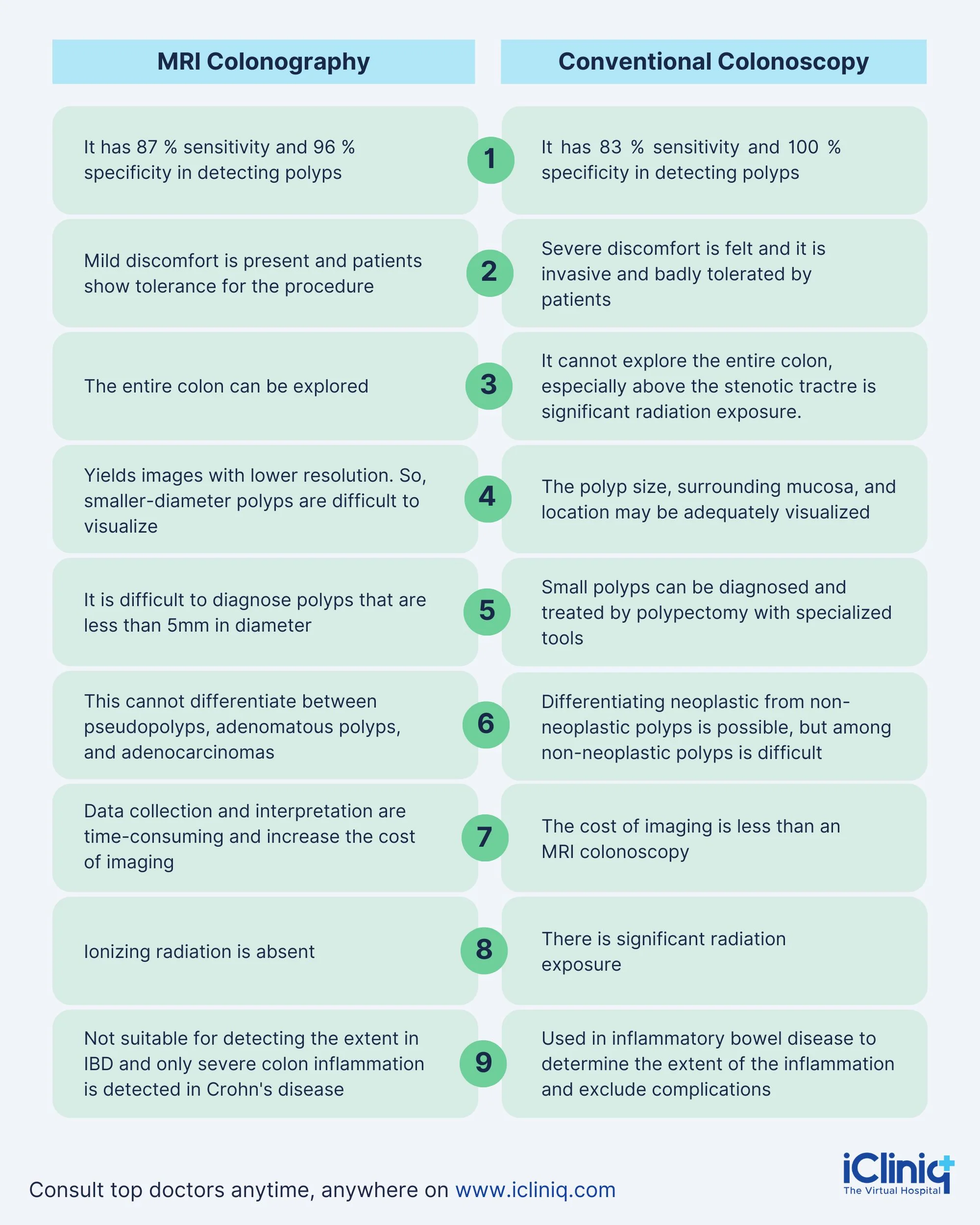
Conclusion:
MRI colonography cannot completely replace conventional colonoscopy in evaluation. The low sensitivity of MRI colonography in mild colonic inflammation limits its use in diseases like ulcerative colitis and Crohn's Disease. Improvements in MRI colonography are under study. They may improve sensitivity for colonic inflammation and spatial resolution.

