Table of Contents
Introduction:
The circulatory system of the human body is complex yet of major significance. It comprises mainly blood vessels that distribute blood throughout the body. Blood vessels are vital nutrient suppliers to tissues, organs, bones, and other key anatomic structures. It mainly comprises arteries and veins. Arteries carry blood from the heart to other tissues, and veins bring them back to the heart. The other important structures of the circulatory system are the lymphatic vessels. They carry lymph, which is a clear fluid containing white blood cells. The lymph plays a vital role in the immunity and defense mechanism of the body, thereby fighting any infections.
What Are Vascular Anomalies?
Vascular anomaly is a broad term used to describe the disorders of the arteries and veins. These arteries, veins, and lymphatic vessels are lined by a special tissue known as the endothelium. Endothelium mainly regulates blood flow and pressure; hence they are significant structures of the blood vessels. Vascular anomalies are abnormalities associated with the endothelium of the blood vessels themselves.
Vascular anomalies are a prevalent condition. They are usually congenital (present at birth). Some anomalies are mild and may cause esthetic problems; however, some could be severe enough to cause functional challenges.
What Are the Types of Vascular Abnormalities?
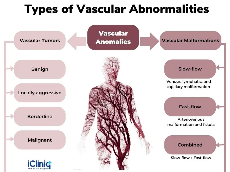
According to the International Society for the Study of Vascular Anomalies (ISSVA), vascular anomalies can be classified into two broad categories as vascular tumors and vascular malformations. Vascular tumors can be further classified as benign (non-cancerous), locally aggressive, borderline, and carcinoma (malignant). In addition, vascular malformations can be classified as slow-flow malformations, fast-flow malformations, and combined malformations.
-
Vascular Tumors:
Vascular tumors are excessive growth or a mass that occurs due to the rapid multiplication of the endothelial cells. Congenital vascular tumors may increase in size after birth. They can be superficial or deep. However, once its complete growth is attained, it may remain stagnant or even shrink in size. Examples include:
-
Benign vascular tumors such as hemangioma and pyogenic granuloma.
-
Malignant tumors like angiosarcoma and Kaposi’s sarcoma.
-
Vascular Malformations:
Vascular malformations refer to the abnormal development of blood vessels. Congenital malformations grow in size and, unlike tumors, do not resolve on their own. They could be present as visible birthmarks requiring cosmetic therapy or might be large enough to cause functional issues. A few examples of vascular malformations are mentioned below:
-
Venous malformations (involvement of veins).
-
Arteriovenous malformations (both arteries and veins are abnormal).
-
Capillary malformations (peripheral vessels are abnormal).
-
Lymphatic malformations (abnormal lymphatic vessels).
What Are the Causes of Vascular Anomalies?
A vascular malformation is a developmental anomaly that occurs during fetal development. Abnormal vascular development could either present as an increased number of endothelial cells or abnormal structural growth of the vessels. Rarely have genetic conditions or hereditary factors known to contribute to the development of vascular malformation.
What Are the Symptoms of Vascular Anomalies?
The signs and symptoms would vary depending on the type of anomaly. They are listed below:
-
Vascular Tumors:
-
Swelling.
-
Tenderness.
-
Bleeding.
-
Recurrent infection or ulceration.
-
Abnormal growth.
-
Vascular Malformation:
-
Pink, red, or purple discoloration of the skin.
-
Swelling.
-
Ulceration.
-
Pain.
-
Impaired movement of the affected region.
-
Facial distortion if the face is affected.
What Is the Role of Imaging Modalities in Diagnosing Vascular Anomalies?
Various imaging modalities can be used to classify and precisely diagnose a vascular anomaly. However, poor image resolution and inappropriate imaging procedures could lead to several diagnostic dilemmas resulting in improper treatment. Hence it is essential to aptly diagnose and characterize the condition for a better patient prognosis. The imaging techniques are described below:
1. Magnetic Resonance Imaging (MRI):
It is the gold standard imaging modality for diagnosing and treating vascular malformations. Gadolinium-based contrast agents are frequently used in MRI examinations. MRI can precisely describe the extent and the hemodynamic nature of the abnormality, whether it is a high or low-flow condition. Contrast MRI is more accurate in characterizing vascular anomalies. MRI can categorize the slow-flow anomalies as lymphatic malformation if no or minimal contrast enhancement is observed and venous malformation if patchy areas are observed. In a malignant tumor, vessels will be randomly distributed within the mass and have an inhomogeneous enhancement.
In contrast, in a benign tumor, vessels are usually located at the periphery and are homogeneous in appearance. However, the major drawback of gadolinium-based contrast agents is the possibility of developing nephrotoxicity. Hence these contrast agents are contraindicated in renal failure and dialysis patients.
2. Magnetic Resonance Angiography (MRA):
MRA is an imaging procedure that captures images of the major blood vessels. This procedure involves injecting a contrast material and imaging through a fluoroscope, computed tomography, or MRI. This is a non-invasive procedure, unlike conventional angiography, where a catheter is used. The advantage of this technique is the images can be obtained from multiple angles, thus creating a more accurate diagnosis.
3. Magnetic Resonance Venography (MRV):
It is used to diagnose low-flow vascular conditions. In addition, it can be used to evaluate the patency of the blood flow in the peripheral veins.
4. Magnetic Resonance Lymphangiography (MRL):
MRL is used in the diagnosis of malignant lymph nodes. This procedure involves injecting the contrast material into the circulation, followed by MRI imaging.
5. Ultrasonography:
Ultrasonography is the fastest and most cost-effective method to diagnose a vascular anomaly. It can be done during the patient’s first visit to the hospital. It is non-invasive, quick, and well-tolerated by patients. Ultrasound uses high-frequency sound waves to capture images of the blood vessels. These sound waves are emitted into the body by a small transducer device. These waves bounce back upon hitting the structures within the body. The transducer captures these returning sound waves and processes them in a computer as images. The computer processes these images based on the velocity, amplitude, and time taken for them to return. A Doppler ultrasound can be used to evaluate blood movement through the vessels and diagnose any mass or obstruction within the vessels. It can also be used to differentiate arterial and venous blood flow.
6. Computed Tomography (CT):
CT is used when MRI is contraindicated, such as when the patient cannot be sedated or has a pacemaker or other medical device. In addition, it is commonly used to determine calcified blood clots and other conditions associated with vascular anomalies.
Conclusion:
Vascular anomalies could result in distressing and apprehensive situations for parents and children. Moreover, misdiagnosis is widespread due to overlapping phenotypic features, which could lead to inappropriate treatment. Hence it is crucial to aptly use imaging modalities for characterizing vascular anomalies, thereby rendering suitable treatment.

